Now that you’ve taken the first step to consult an endometriosis specialist regarding your endometriosis symptoms, what’s next in this journey? One of the first and most important things that your gynaecologist does would be to assess how severe your endometriosis is, in a process known as endometriosis staging. This staging procedure will help guide your gynae in the creation of your treatment plan, and provide more insight on the outcomes of treatment.
Endometriosis Staging
Endometriosis has often been staged according to the revised American Fertility Society (rAFS) scoring system, which grades endometriosis into stages 1, 2, 3, 4, with stage 4 being the most advanced, with severe adhesions involving the ovaries, fallopian tubes and organs such as the rectum.
Stage of Endometriosis |
Measurement of Endometriosis |
Stage 1: Minimal |
There are some small lesions and endometrial tissue growing on the ovaries. Some inflammation around the pelvic area might be observed. |
Stage 2: Mild |
There are small lesions and shallow implants on the ovaries and pelvic area. |
Stage 3: Moderate |
There are more lesions and deep implants on the ovaries and pelvic area. |
Stage 4: Severe |
There are even more lesions and deeper implants on the ovaries and pelvic area. Other organs such as the bladder, ureter and bowels may also be affected. |
Table Explaining the Different Stages of Endometriosis
Traditionally, the rAFS scoring is performed surgically as the gold standard for the diagnosis of endometriosis was by laparoscopy. In this procedure, a telescope is inserted through the belly button into the abdominal cavity for visual exploration. With magnification and the ability to move and bring the laparoscope close to organs and structures of interest, the gynaecologist can then confirm the sites and severity of the endometriosis.
However, laparoscopic diagnosis also meant that your gynaecologist would only be able to tell you how bad the endometriosis was after the surgery, rather than prior to it. Thus, in the early years of endometriosis treatment, there were probably quite a bit of under-diagnosis cases due to diagnostic limitations, if surgery was not performed.
The other key drawback of this conventional approach is that the operating gynaecologist may not be prepared to tackle and remove as much of the endometriosis as possible especially if it was severe, since there was no opportunity prior to surgery to discuss the extent of the surgery required. In severe cases of endometriosis, other specialties e.g. urologists or colorectal surgeons may need to be involved, especially when radical excision of endometriosis is needed.
An Advancement in Endometriosis Staging – The Power of Ultrasound for Endometriosis Mapping
 In 2016, the International Deep Endometriosis Analysis (IDEA) group published a consensus opinion on sonographic features (features that can be observed with ultrasound) of the different types of endometriosis. This gave rise to the concept of endometriosis mapping and its use for prediction of the complexity of surgery required for the patient.
What is Endometriosis Mapping?
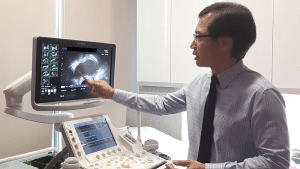
Endometriosis mapping is a systematic and thorough ultrasound examination of the different structures and organs in the pelvis, and assesses whether their normal function and organizational relationship to each other is being affected by endometriosis. In doing so, it is able to “stage” the disease and help predict the complexity of treatment, whether by surgery or medical treatment.

Benefits of Mapping with Ultrasound
The importance of Endometriosis mapping with transvaginal ultrasound is that it gives a “window” into the severity of the disease, and allows differentiation into mild/moderate/severe disease. The Endometriosis specialist has a chance to coordinate care accordingly, involving fertility specialists, colorectal specialists and also urological specialists. The current concept in top centres treating endometriosis is to minimize surgery and maximize fertility outcomes for women who plan to have children. Without knowing the full extent of the disease, it is almost impossible to achieve this.
Although MRI is another modality to assess the severity of endometriosis — it has excellent performance characteristics in differentiating soft tissue and deep endometriosis and is superior to CT scan — it is expensive and not so readily available in many countries.

The accuracy of transvaginal ultrasound in assessment of endometriosis has been validated in many studies, with accuracy as high as 94% for moderate and severe pelvic endometriosis. A transvaginal ultrasound can be performed at the doctor’s office with minimal preparation, at an affordable price without the need to head to an imaging centre. All that is required is a gynae who is experienced in performing such ultrasound scans.
Here’s a quick recap on the various staging methods used to determine the severity of endometriosis:
Transvaginal Ultrasound |
MRI |
CT scan |
|
Cost |
$ |
$$$ |
$$ |
Convenience |
🙂🙂🙂 |
🙂 |
🙂 |
Dynamic/ Static Imaging |
Dynamic (able to test the mobility and relationship of organs to each other, doctor can also assess for pain and elasticity of tissues at same time) |
Static (the organs are in a resting state hence not able to test the mobility of organs, doctor is unable to interact with patient during procedure) |
Static |
Dependency on skill of doctor/radiographer |
🙂🙂🙂 |
🙂 |
🙂 |
Accuracy |
🙂🙂🙂
Able to detect small endometriosis lesions eg 1cm DIE |
🙂🙂🙂
Able to detect small endometriosis lesions. |
🙂 Offers some degree of detection |
Radiation exposure |
Nil |
Nil |
Yes |
Coverage of assessed area |
Smaller, within pelvis only |
Wider area |
Wider area |
Conclusion
While laparoscopic surgery is the gold standard in the diagnosis and confirmation of endometriosis and staging, it is an invasive procedure and provides details only after a surgery is done. In order to reduce the number of disruptive surgeries, it will be much more useful to know the stage of endometriosis involvement even before planning for any surgery, so that a less-is-more approach can be adopted.
- Part 1: The Different Faces of Endometriosis
- Part 2: What Your Ultrasound Scan Says About Your Endometriosis
- Part 3: Why Do Endometriosis Symptoms Continue After Treatment
- Part 4: Your Unique Endometriosis Map – An Important Tool for Treatment


